发布时间:2026-10-16 17:15:50 来源: 浏览次数:104
《International Journal of Hyperthermia》
2026, VOL. 43, NO. 1, 2612543
Polidocanol augments high-intensity focused ultrasound efficacy for hyperintense fibroids on T2WI: a retrospective cohort study
Ming-tao Yang, Ling Tian, Yu Xia, Hong-ni He, Chuan Lin, Tian-wen He, Ying Tang , Hui-quan Hu and Jun Li
Department of Obstetrics and Gynecology, The Second Clinical Medical College of North Sichuan Medical College, Nanchong, Sichuan, China
ABSTRACT:
Objective: To compare the immediate efficacy and safety of high-intensity focused ultrasound (HIFU) monotherapy with the combination of ultrasound-guided intratumoral polidocanol injection and HIFU for the treatment of uterine fibroids with highT2-weighted imaging (T2WI) signal intensity.
Methods: In this retrospective study, 116 patients with T2-hyperintense uterine fibroids treated between January 2024 and May 2025 were enrolled. Patients were allocated to either HIFU monotherapy (the H group, n =63) or combined polidocanol and HIFU therapy (the H +P group, n =53). Thereafter, therapeutic parameters, the non-perfused volume ratio (NPVR), and adverse events were compared between the two groups after propensity score matching.
Results: The matched cohort comprised 53 patients per group. The median NPVR was significantly higher in the H +P group than in the H group (92.57% vs. 77.81%, p <0.001). Additionally, a significantly greater proportion of patients in the H +P group achieved an NPVR ≥ 80% (87% vs. 43%, p <0.001) and an NPVR ≥ 90% (60% vs. 9%, p <0.001). The H +P group also required significantly less sonication time and lower total energy delivery (both p <0.05). Furthermore, the overall incidence of adverse events was similar between the two groups (73.6% vs. 69.8%, p =0.506). However, the H +P group exhibited a markedly lower incidence of sacrococcygeal pain (5.7% vs. 45.3%, p <0.001).
Conclusion: Pretreatment with ultrasound-guided intratumoral polidocanol injection may improve the immediate ablation efficacy of HIFU in T2-hyperintense uterine fibroids, reducing energy requirements and mitigating procedure-related sacrococcygeal pain. These findings highlight the potential of this combined therapy and underscore the need for prospective studies to confirm its long-term clinical benefits.
KEYWORDS High-intensity focused ultrasound; polidocanol;hyperintense fibroids on T2WI; uterine fibroids;efficacy
Introduction
Uterine fibroids are the most common benign tumors in women of reproductive age, with a prevalence rate of 20%–77% [1]. Approximately 30%-50% of patients present with symptoms such as menorrhagia, pelvic pain, and frequent urination, significantly impairing their quality of life [2].
High-intensity focused ultrasound (HIFU), as a noninvasive thermal ablation technique, induces tumor necrosis through the precisely focused ultrasound waves, which offers advantages such as minimal trauma and rapid recovery [3]. It has become a preferred treatment option for patients desiring fertility preservation or those seeking to avoid surgery [4]. HIFU works by concentrating ultrasonic energy on the target lesion, causing coagulative necrosis in the fibroid tissue. However, the characteristics of the target medium may affect ultrasound energy deposition, thereby affecting the therapeutic efficacy. Recent studies have highlighted that MRI T2-weighted imaging (T2WI) signal intensity is the most critical influencing factor for ablation efficiency [5]. Fibroids with high T2 signal intensity posesignificantly greater clinical challenges than those with low or isointense signals [6]. Their tissue properties impede energy deposition, limiting the HIFU ablation efficacy, often resulting in insufficient nonperfused volume (NPV) ratios (NPVRs) [7,8]. These limitations may hinder symptom relief and necessitate secondary interventions. Consequently, optimizing protocols for hyperintense fibroids on T2WI has become a research priority.
Recent studies have suggested that combination therapies may enhance the HIFU efficacy for refractory fibroids [9–11]. Polidocanol, as a sclerosing agent, reduces lesion blood supply through vascular embolization and has been safely used in various conditions [12,13]. Ultrasound-guided Polidocanol-assisted radiofrequency ablation (RFA) shows clinical value for vascular subserosal fibroids [14]. Notably, when mixed with air to form foam, Polidocanol may theoretically enhance cavitation and thermal effects during HIFU [9,10,15]. However, clinical evidence regarding its efficacy specifically for fibroids with high T2 signals remains limited.
Based on these theoretical advantages, our institution has implemented the combined Polidocanol-HIFU therapy for selected hyperintense fibroids on T2WI. This retrospective study aimed to compare HIFU monotherapy with the combination of Polidocanol pretreatment and HIFU in this challenging subgroup, and evaluate their immediate efficacy and safety through historical treatment data.
Materials and methods
Study design and setting
This single-center retrospective cohort study was conducted in accordance with the Strengthening the Reporting of Observational Studies in Epidemiology (STROBE) guidelines. Historical treatment data of patients with uterine fibroids treated at our institution between January 2024 and May 2025 were collected. Through systematic review of electronic medical records, 116 consecutive patients meeting the following criteria were identified. Inclusion criteria: (1) reproductive-age women; (2) those showing documented preference for uterine preservation; (3) having single fibroid with high T2 signal intensity on magnetic resonance imaging (MRI) (defined as signal intensity >myometrium [17]); and (4) with complete pre- and post-treatment imaging data. Exclusion criteria: (1) pregnant or breastfeeding women (verified by β-human chorionic gonadotropin testing); (2) those with active gynecological pathologies (e.g. histological-confirmed malignancies, acute pelvic inflammatory disease (PID)); (3) connective tissue disorders (systemic lupus, scleroderma, etc.); (4) MRI evidence of fibroid degeneration (necrosis > 50%, calcification, red degeneration); (5) recent (< 6 months) thromboembolic or hemorrhagic events; and (6) severe organ dysfunction (estimated glomerular filtration rate (eGFR) < 30, and left ventricular ejection fraction (LVEF) < 40%, etc.). The Ethics Committee of North Sichuan Medical College granted exemption from informed consent (IRB:2025120), because only deidentified historical data were analyzed, in compliance with the Declaration of Helsinki principles.
Data collection and variables
Data were extracted from electronic medical records by two independent investigators according to a standardized protocol. Variables collected included demographics: age and body mass index (BMI); fibroid characteristics: volume (cm³), location (submucosal, intramural, subserosal), and position (anterior, posterior, lateral, fundal); MRI parameters: subcutaneous fat thickness (mm), fibroid-sacrococcyx distance (mm), and T2WI signal intensity (quantitative region of interest (ROI) analysis); ultrasound parameters: blood flow grade (0–4 scale); treatment parameters: sonication time, total energy, and treatment intensity. Our outcomes were NPVR and adverse events. Based on the actual treatments received, patients were divided into the H group (n =63) receiving HIFU monotherapy, and the H +P group (n =53) undergoing ultrasound-guided intratumoral polidocanol injection combined with HIFU.
Pre-HIFU MRI and ultrasound evaluation
MRI examination was performed using the GE Signa HDxt 1.5 T superconducting MRI scanner. Patients underwent MRI scans within three days before HIFU treatment according to the standardized protocol. Based on the Funak criteria, fibroids were classified as hypointense, isointense, or hyperintense according to the T2WI signal intensity [16]. Hyperintense fibroids on T2WI were defined as those with signal intensity values exceeding that of the myometrium [17]. All patients underwent transvaginal ultrasound(including color Doppler imaging) upon admission, and the fibroids were semi-quantitatively graded based on blood supply in line with the following criteria: grade 0, no blood flow signal; grade 1, minimal blood flow with 1–2 punctate signals; grade 2, moderate blood flow with one main vessel longer than the fibroid radius or 2–3 small vessels; grade 3, abundant blood flow with more than four interconnected vessels; and grade 4, extremely abundant blood flow with more than four reticular interwoven vessels or vessel diameter exceeding 3 mm.
Pretreatment preparation
Patients in both the H and H +P groups completed laboratory tests to confirm their compliance with inclusion and exclusion criteria. They were required to follow a liquid diet two days preoperatively and take laxatives with enema administration one day before the procedure. On the day of treatment, the lower abdomen underwent skin preparation, degreasing, and degassing, followed by urinary catheter placement. The anesthesia protocol was as follows: based on patient weight, fentanyl and midazolam were administered at the initial intravenous doses of 1 μg/kg and 0.03 mg/kg, separately followed by supplemental doses of 0.8 μg/kg and 0.02 mg/kg, respectively, after 30 min. Subsequent doses were repeated at 30-min intervals at the same dosage. Throughout the procedure, patients tolerated treatment discomfort, maintained normal cardiopulmonary function, and responded appropriately to verbal and light tactile stimuli.
Treatment process
The treatment was performed using the JC200 HIFU system (Chongqing Haifu Medical Technology Co., Ltd., Chongqing, China) for HIFU therapy. The system was integrated with an ultrasound imaging device(MyLab 70, Esaote, Italy) to enable real-time treatment guidance. All procedures were accomplished in the ultrasound scanning mode with a slice spacing of 0.5 mm. The parameters of the ultrasound transducer were shown below: frequency of 0.8 MHz, diameter of 200 mm, focal length of 155 mm, power range of 0–400 W, and physical focal zone of 1.5 × 1.5 × 10 mm. Patients in the H group were placed in a prone position on the HIFU treatment bed, and electrocardiographic monitoring and intravenous access were established. Before HIFU treatment, real-time ultrasound was carried out to localize the fibroid, ensuring a clear acoustic pathway and avoiding intestinal injury. A treatment plan was subsequently formulated with the power being set at 400 W in the point-scanning mode. Treatment completion was determined when the target fibroid area exhibited a hyperechoic patch that covered the entire fibroid or significant grayscale enhancement.
For patients in H +P group, ultrasound-guided polidocanol foam injection was performed prior to HIFU ablation. The specific procedure was as follows. For sclerosant preparation, a 1% polidocanol solution (Xi’an Tianyu Pharmaceutical Co., Ltd., China) was used. The polidocanol dosage was calculated using the formula V =4/3π(r+0.5)³, where V represents the total dose and r denotes the lesion radius in centimeters. To produce the foam, half of the calculated polidocanol solution was mixed with air at a 1:1 ratio. The Tessari method was employed to prepare the sclerosing foam. To be specific, a 10 ml syringe containing 1% polidocanol solution was connected to a 10 ml syringe containing an equal volume of room air via a three-way stopcock, achieving a liquid-to-air ratio of 1:1 . Thereafter, these two syringes were rapidly alternated more than 20 times to form a uniform, fine-textured foam. The prepared foam was highly stable and injected within 90 s to ensure the optimal efficacy. With regard to the injection technique, under continuous real-time ultrasound guidance, a 17 G PTC needle was percutaneously or transvaginally inserted into the target area of the uterine fibroid. Notably, the puncture path was carefully planned to avoid critical structures such as the intestines and major blood vessels. Afterwards, based on pretreatment color Doppler ultrasound findings, the needle tip was positioned within the peripheral region of the lesion and/or areas with abundant blood flow signals. For site distribution and injection dosage, a fan-shaped distribution strategy was adopted to administer polidocanol and polidocanol foam at multiple sites within the fibroid, ensuring extensive coverage of the vascular network. The injection volume was adjusted according to the agent diffusion range within the fibroid. The maximum foam volume per treatment session was set at 15 ml, and the maximum drug dose should not exceed 30 ml. Aspiration was performed before each injection, so as to confirm that the needle tip was not inside a blood vessel. During perioperative monitoring, patients were closely monitored throughout the injection procedure for adverse reactions, including but not limited to changes in heart rate, blood pressure, oxygen saturation, and signs of allergic reactions. Emergency medications and equipment were readily available. Within 1-h postpolidocanol injection, the H +P group underwent HIFU treatment identical to that administered to the H group. Parameters including HIFU treatment duration, procedural time, treatment intensity, and energy dose were recorded for patients in the two groups.
Post-HIFU procedure evaluation
All patients underwent immediate on-board ultrasound measurement of the three-dimensional diameters of the fibroid (longitudinal diameter (d1), longitudinal diameter (d2), and transverse diameter (d3)) following HIFU treatment. Over 5 min after HIFU completion, 1.5 ml of reconstituted SonoVue (each vial containing 25 mg of SonoVue, Bracco, Milan, Italy, which was mixed with 5 ml of saline) was administered intravenously. Two min later, the NPV was assessed and measured through on-board ultrasound. The volumes of both the fibroid and the NPV were calculated using the formula V = (d1 ×d2 ×d3) × 0.5233. The primary efficacy endpoint was the non-perfused volume ratio (NPVR%), which was calculated by(non-perfused volume/initial fibroid volume) × 100%. To reflect the clinical relevance, the primary binary outcome was defined as the proportion of patients achieving technical success (an NPVR ≥ 80%). Whereas a more stringent secondary outcome was set as the proportion of patients achieving an NPVR ≥ 90%. Success rate refers to the proportion of patients immediately achieving the predefined technical success criteria (NPVR ≥ 80%) or the more stringent secondary criteria (NPVR ≥ 90%) post-treatment.
Evaluation of side effects
The maximum VAS Pain score (0–10) and skin assessment were documented in nursing records. According to the Society of Interventional Radiology procedure, side effects were classified as grades A–F [9]. All adverse events were monitored and then recorded during the HIFU procedure and throughout the postoperative inpatient observation period. After the treatment, patients were monitored in the HIFU monitoring ward for at least 24 h and discharged only after being assessed as stable. As a result, the adverse events reported in this study reflect the short-term safety profile in the perioperative period.
In addition, this study specifically monitored the potential adverse reactions directly associated with the polidocanol injection procedure in the combination therapy group (H +P group), covering the period from the start of the injection until immediately before the subsequent HIFU treatment. The monitoring focused on allergic reactions (such as rash and dyspnea), cardiovascular responses(including significant fluctuations in heart rate or blood pressure), and potential complications theoretically associated with inadvertent intravascular injection.
Statistical analysis
SPSS 27.0 software (SPSS Inc., Chicago, IL, USA) and R software (version 4.2.2) were employed for statistical analyses. Categorical variables were expressed as frequencies, and between-group comparisons for binary outcomes were presented as n/N (%). Continuous variables were described as median(interquartile range, IQR) or mean ±standard deviation. Normality of data was first assessed using the Kolmogorov-Smirnov test. Then, normally distributed quantitative variables were analyzed by t-tests, while non-normally distributed variables were analyzed with the Mann-Whitney U test. For between-group comparisons, categorical variables were evaluated by chi-square tests or Fisher’s exact probability test (by Yates-corrected χ2 test when all the expected frequencies were ≥ 5; and by Fisher’s exact test at the expected frequencies < 5). A logistic regression model was utilized to identify factors influencing an NPVR ≥ 80% and sacrococcygeal pain. Moreover, propensity scores were calculated through logistic regression based on demographic and clinical characteristics, followed by 1:1 nearest neighbor matching using the MatchIt package in R to minimize the total absolute distance across the matched pairs, with a caliper value of 0.2. This ensured covariate balance between the matched groups (all the standardized differences < 10%). A P-value < 0.05 was considered statistically significant.
Results
Patient characteristics
A total of 116 patients were enrolled for analysis, including 63 in the H group and 53 in the H +P group. Following propensity score matching, a total of 106 patients with fibroids were included in the matched cohort, including 53 (50%) in the H group and 53 (50%) in the H +P group. Figure 1 shows the patient flow diagram. Differences in age, BMI, uterine position, rectus abdominis muscle thickness, subcutaneous fat layer thickness, distance from the posterior aspect of fibroids to the sacrococcyx, ultrasound blood flow grading, fibroid type, fibroid location, and fibroid volume did not show any statistical significance between the two groups (p>0.05, Table 1).
Treatment parameters
All patients successfully completed the ultrasound ablation therapy. The median sonication time in the H +P group was significantly shorter than that in the H group (578 s [IQR: 399–745] vs. 758 s [IQR: 408–1128]; p =0.022). The median total energy delivered was also significantly lower in the H +P group(226,800 J [IQR: 157,560–298,200] vs. 303,200 J [IQR: 163,200–451,200]; p =0.013). However, there was no significant difference in the total operation time or treatment intensity between the two groups (p>0.05, Table 2).
Comparison of NPV outcomes
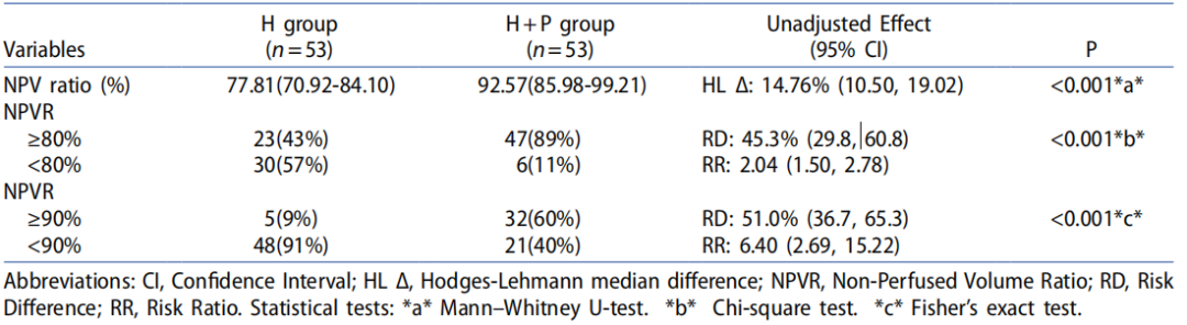
The H +P group showed significantly superior NPV outcomes to the H group. To be specific, the median NPVR was higher in the H +P group (92.57% vs 77.81%, p <0.001). With regard to the primary binary outcome (an NPVR ≥ 80%), the success rates were 89% in the H +P group versus 43% in the H group(p <0.001). For the secondary outcome (an NPVR ≥ 90%), the success rates were 60% in the H +P group versus 9% in the H group (p <0.001, Table 3).
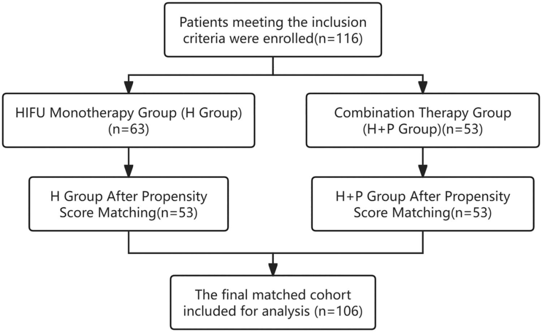
Figure 1. patient enrollment flowchart.
Table 1. patient demographics and baseline characteristics.
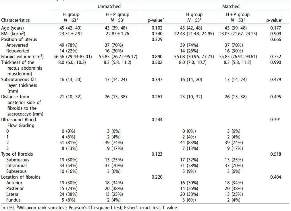
Table 2. Comparison of therapeutic parameters between H group and H + p group

Table 3. Comparison of NPV rates between the two groups of patients.
Univariate and multivariate analyses of factors influencing an NPVR ≥ 80%
In the univariate analysis, treatment modality was the only factor significantly associated with an NPVR ≥ 80%. Compared with patients receiving HIFU alone, those who received combined HIFU and polidocanol treatment were significantly more likely to achieve an NPVR ≥ 80% (OR = 8.57, 95% CI: 3.27–22.45, p <0.001). Other factors, including age, BMI, uterine position, ultrasound blood flow grading, type of fibroids, location of fibroids, and fibroid volume, were not significantly associated with the outcome. In the multivariate analysis, after adjusting for all the included covariates, the HIFU +polidocanol treatment remained a strong and independent predictor of an NPVR ≥ 80% (adjusted OR = 15.13, 95% CI: 4.66–49.12, p <0.001, Table 4).
Table 4. univariate and multivariate analysis of influencing NPVR ≥80% factors.
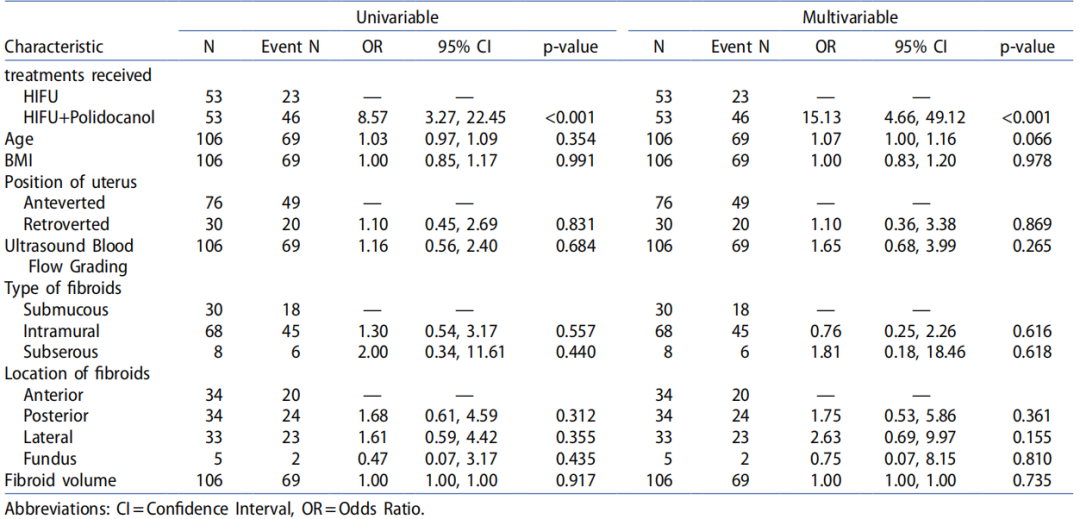
Table 5. Incidence rate of adverse effect during HIFU treatment.
Safety of polidocanol injection
Throughout the H +P group (n =53), no adverse reactions directly attributable to polidocanol injection were reported during the monitored interval (from the start of injection until immediately before HIFU treatment).
Adverse events (AEs) during HIFU treatment
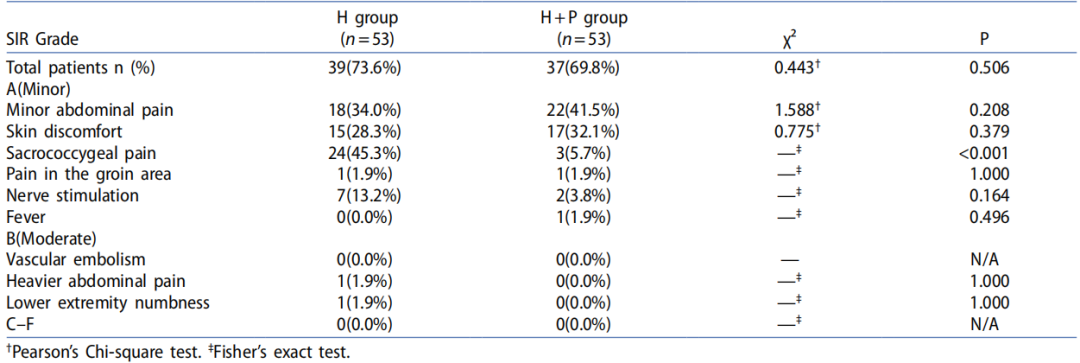
The incidence and severity of AEs during HIFU treatment are summarized in Table 5. As observed, the overall incidence of AEs was comparable between the H group (73.6%) and the H +P group (69.8%)(p =0.506). The profile of minor AEs (SIR Grade A), including abdominal pain, skin discomfort, pain in the groin area, nerve stimulation, and fever, was largely similar between the two groups (p>0.05). However, there was a statistically significant difference in the incidence of sacrococcygeal pain, which was reported in 24 patients (45.3%) in the H group whereas only in 3 patients (5.7%) in the H +P group (p <0.001). Moderate AEs (SIR Grade B) were rare. One case of heavier abdominal pain and lower extremity numbness was reported in the H group, while such events were not reported in the H +P group, and the differences were not statistically significant (p = 1.000). No severe AEs (SIR Grades C-F) or vascular embolism events occurred in either group.
Table 6. univariate and multivariate analysis of influencing sacrococcygeal pain factors.
Risk factors for sacrococcygeal pain
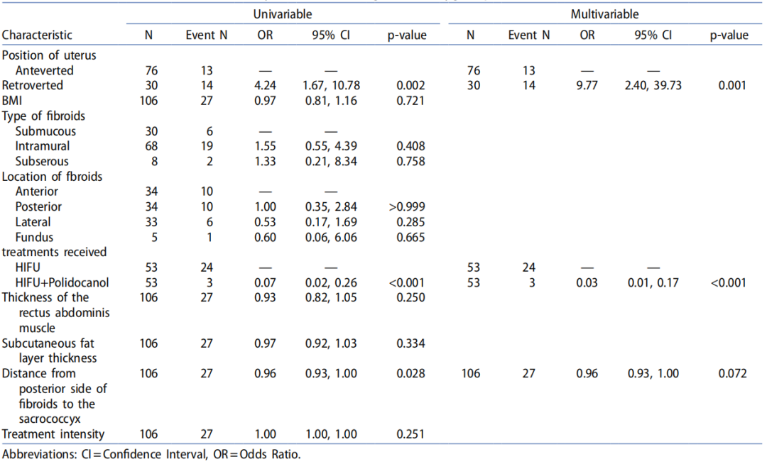
In univariate analysis, retroverted uterus (OR = 4.24, 95% CI: 1.67–10.78, p =0.002), treatment with HIFU alone (reference: HIFU +polidocanol; OR = 0.07, 95% CI: 0.02–0.26, p <0.001), and a shorter distance from the fibroid to the sacrococcyx (OR = 0.96, 95% CI: 0.93–1.00, p =0.028) were the significant risk factors for sacrococcygeal pain. In the multivariate model, after adjusting for confounding variables, both retroverted uterus and treatment type remained the independent and significant predictors of sacrococcygeal pain. Moreover, the odds of the event was substantially higher for patients with a retroverted uterus(adjusted OR = 9.77, 95% CI: 2.40–39.73, p =0.001). Conversely, the HIFU +polidocanol treatment remained a strong protective factor (adjusted OR = 0.03, 95% CI: 0.01–0.17, p <0.001, Table 6).
Discussion
This retrospective analysis suggested that the combination of intratumoral polidocanol injection and HIFU might enhance the immediate treatment efficacy for T2-hyperintense uterine fibroids. The observed superior NPVR and reduced energy requirements in the H +P group warrant further investigation. Notably, the H +P group demonstrated a lower incidence of sacrococcygeal pain, though retrospective data preclude causal attribution.
NPVR serves as a critical predictor for HIFU outcomes. Prior evidence has indicated that the NPVR >80% is correlated with improved volume reduction and reduced reintervention rates [5,8]. Prior to HIFU ablation, MRI has become an essential tool for predicting and assessing the fibroid treatment efficacy, with T2WI signal intensity being the most pivotal indicator of HIFU effectiveness [3,18–20]. Due to their higher cellularity, lower collagen content, abundant fluid, and heightened vascularization, these features impede ultrasonic energy deposition [3,11,19]. A retrospective study involving 339 patients reported a median NPVR of 75.2% (IQR: 31.5%) for hyperintense fibroids [21]. Our findings aligned with these results: among the 53 patients with T2WI hyperintense fibroids treated with HIFU alone, the median NPVR was77.8%, suggesting that it remains challenging to achieve an NPVR of 80% with the current HIFU technology for such fibroids.
Multiple studies have demonstrated that sulfur hexafluoride microbubbles can safely enhance and accelerate HIFU ablation for uterine fibroids [22,23]. Research exploring the feasibility of combining HIFU with ultrasound-guided intratumoral ethanol injection has reported improved ablation rates, shorter procedure times, lower energy doses, and significant reductions in patient pain and side effects[9]. However, none of these studies address the outcomes for T2WI hyperintense fibroids. As suggested in a propensity score-matched cohort study, GnRH-a pretreatment combined with HIFU may improve the efficacy for heterogeneous T2WI hyperintense fibroids [11,21]. Nevertheless, the GnRH-a combination therapy presents drawbacks, where the insufficient estrogen levels may trigger severe perimenopausal symptoms, leading to treatment discontinuation and compromised efficacy [21]. Additionally, this regimen requires a 3-cycle GnRH-a pretreatment, prolonging the overall treatment duration [11]. Thus, it is imperative to explore novel strategies to improve the NPVR for T2WI hyperintense fibroids. Our study demonstrated that the combined therapy group achieved a significantly higher NPVR (92.57%), exceeding the efficacy threshold of 80% in 89.8% of cases. While retrospective studies suggest that increases in NPVRs may reduce the recurrence risk [7], our findings require prospective validation regarding the long-term symptom relief. The mechanism underlying the improved ablation efficiency may be that, polidocanol foam microbubbles potentially lower the cavitation threshold, facilitating energy deposition at the lower acoustic power [10,24]. Sclerosant-induced vascular occlusion may reduce the heat-sink effects [12,14].
Uterine fibroids are a key focus in obstetrics and gynecology, and current research has emphasized minimally invasive treatments. Patients with T2 hyperintense fibroids may benefit from this combined therapy, which offers a minimally invasive option while reducing drug-related adverse effects (e.g. menopausal symptoms and decreased bone density). In our study, the comparable overall adverse reaction rates (73.6% vs 69.8%) suggested no major safety concerns. The significantly lower sacrococcygeal pain incidence in the H +P group (5.7% vs 45.3%) merits attention, though confounding factors (like patient individual variation) cannot be excluded. Vascular complications theoretically associated with sclerosants were not observed, possibly due to localized administration [13]. Moreover, the absence of injection-related adverse events in the H +P group suggests that, under the study protocol utilizing ultrasound-guided precise intralesional injection, polidocanol administration itself exhibits a favorable procedural safety profile.
While the findings are encouraging, several limitations in this study should be noted. First, although propensity score matching and multivariate adjustments were employed in this retrospective non-randomized study to adjust for confounders, there are still risks of selection bias and unmeasured confounding due to its inherent nature. Treatment allocation was based on clinical judgment, and residual confounding (such as subtle variations in the fibroid texture or patient anatomy not fully captured by the variables) may persist, even though key baseline characteristics were balanced post-matching. Second, fibroid characterization was dependent on the routine clinical MRI sequences. Considering the absence of quantitative T2-weighted imaging analysis, it is impossible to further investigate the relationship between specific signal intensity values and ablation efficacy. Furthermore, although perioperative safety profiles were favorable, the follow-up period was limited to short-term outcomes. The lack of systematic long-term data, including fibroid volume reduction, symptomatic improvement, and recurrence rates, constrains our understanding of the sustained clinical benefits of the combined therapy. Given the above-mentioned limitations and the preliminary evidence gathered, future studies should prioritize large-scale prospective randomized controlled trials. Incorporating quantitative MRI parameters, such as T2 mapping and perfusion metrics, would be invaluable for establishing reliable predictive biomarkers for treatment success. Additionally, subsequent efforts should emphasize long-term follow-up to assess the patient-centered outcomes, including quality of life, degree of symptom relief, and need for reintervention. Besides, complementary laboratory studies exploring the optimal dosage of polidocanoland its underlying biophysical mechanisms can further refine the treatment protocol and enhance our understanding of how sclerosants augment HIFU ablation.
Conclusion
Retrospective data indicate that ultrasound-guided intratumoral polidocanol combined with HIFU may potentially improve the immediate ablation efficiency in high-T2 signal uterine fibroids, with favorable energy utilization and pain outcomes. Prospective studies are needed to establish the long-term clinical benefits.
Author contributions
This research was developed by Ming-tao Yang, Hui-quan Hu, Jun Li, and Hong-ni He, and carried out by Chuan Lin, Yu Xia and Ling Tian. Ming-tao Yang and Tian-wen He contributed information from medical records. Jun Li assisted all authors in drafting the original manuscript. The article’s final form was written and approved by all of the writers.
Disclosure statement
No potential conflict of interest was reported by the author(s).
Funding
This work was supported by the Project of Nanchong Science and Technology Bureau [Grant number. 20SXQT0002],the Foundation of State Key Laboratory of Ultrasound in Medicine and Engineering [Grant number. 2020KFKT003,2021KFKT022] and the North Sichuan Medical College Youth Program under Grant [Grant number. CBY23-QNA19].
ORCID
Ying Tang
http://orcid.org/0000-0001-9318-8986
Data availability statement
The datasets generated and analyzed in the current study are available from the corresponding author.
References
[1] Giuliani E, As-Sanie S, Marsh EE. Epidemiology and management of uterine fibroids. Int J Gynaecol Obstet.2020;149(1):3–9 . doi: 10.1002/ijgo.13102 .
[2] Management of Symptomatic Uterine Leiomyomas: ACOG Practice Bulletin, Number 228. Obstet Gynecol.2021;137(6):e100–e115 .
[3] Wang L, Liu Y, Lin J, et al. Effect of "T2-rim sign" related parameters on high-intensity focused ultrasound ablation of uterine fibroids. Eur J Radiol. 2024;181:111767 . doi: 10.1016/j.ejrad.2024.111767 .
[4] Li W, Jiang Z, Deng X, et al. Long-term follow-up outcome and reintervention analysis of ultrasound-guided high intensity focused ultrasound treatment for uterine fibroids. Int J Hyperthermia. 2020;37(1):1046–1051 . doi:10.1080/02656736 .2020.1807617.
[5] Yang MJ, Yu RQ, Chen WZ, et al. A prediction of NPVR ≥ 80% of ultrasound-guided high-intensity focused ultrasound ablation for uterine fibroids. Front Surg. 2021;8:663128 . doi: 10.3389/fsurg.2021.663128 .
[6] Funaki K, Sawada K, Maeda F, et al. Subjective effect of magnetic resonance-guided focused ultrasound surgery for uterine fibroids. J Obstet Gynaecol Res. 2007;33(6):834–839 . doi: 10.1111/j.1447-0756.2007.00665.x.
[7] Dou Y, Zhang L, Liu Y, et al. Long-term outcome and risk factors of reintervention after high intensity focused ultrasound ablation for uterine fibroids: a systematic review and meta-analysis. Int J Hyperthermia.2024;41(1):2299479 . doi: 10.1080/02656736 .2023.2299479.
[8] Yang M, Chen Y, Zhou X, et al. Machine learning models for prediction of NPVR ≥80% with HIFU ablation for uterine fibroids. Int J Hyperthermia. 2025;42(1):2473754 . doi: 10.1080/02656736.2025.2473754 .
[9] Yang Z, Zhang Y, Zhang R, et al. A case‐control study of high‐intensity focused ultrasound combined with sonographically guided intratumoral ethanol injection in the treatment of uterine fibroids. J Ultrasound Med.2014;33(4):657–665 . doi: 10.7863/ultra.33.4.657.
[10] Farny CH, Glynn Holt R, Roy RA. The correlation between bubble-enhanced HIFU heating and cavitation power. IEEE Trans Biomed Eng. 2010;57(1):175–184 . doi: 10.1109/TBME.2009.2028133 .
[11] Yang S, Kong F, Hou R, et al. Ultrasound guided high-intensity focused ultrasound combined with gonadotropin releasing hormone analogue (GnRHa) ablating uterine leiomyoma with homogeneous hyperintensity on T (2) weighted MR imaging. Br J Radiol. 2017;90(1073):20160760 . doi: 10.1259/bjr.20160760 .
[12] Min X, Zhang Z, Chen Y, et al. Comparison of the effectiveness of lauromacrogol injection for ablation and microwave ablation in the treatment of predominantly cystic thyroid nodules: a multicentre study. BMC Cancer.2023;23(1):785 . doi: 10.1186/s12885-023-11301-7 .
[13] Qi Y, Huang Y. Treatment for abdominal wall endometriosis: ultrasound-guided percutaneous injection of lauromacrogol sclerotherapy. J Minim Invasive Gynecol. 2023;30(3):171–172 . doi: 10.1016/j.jmig.2022.12.009.
[14] Qi YF, Dong XQ, Zhang LW, et al. Clinical value of ultrasound-guided lauromacrogol-assisted radiofrequency ablation for uterine fibroids. Chinese Journal of Ultrasonography. 2016;32(11):1022–1024 . Retrieved from: https:// kns.cnki.net/kcms2/article/abstract v=yqBhao7Q9gxXndGPYvbNr7ihIZuCi-iEZT We-KcWY12_nuCPyPhRdsgV Xn3mB v ni2iST FW oL k uo yj m25Cb qx9JIG bKa E ep9J ZD2FT OrjG ll wsj Dd60tV L c d ks mm8Ygj9c2dZKJpH3zlRAGDn2d0UtIMBABKFxIsZgq-77jpUD0oWW29jUUlcg==&uniplatform=NZKPT&language=CHS
[15] Chen Y, Jiang J, Zeng Y, et al. Effects of a microbubble ultrasound contrast agent on high-intensity focused ultrasound for uterine fibroids: a randomised controlled trial. Int J Hyperthermia. 2018;34(8):1311–1315 . doi:10.1080/02656736 .2017.1411620.
[16] Funaki K, Fukunishi H, Funaki T, et al. Magnetic resonance-guided focused ultrasound surgery for uterine fibroids: relationship between the therapeutic effects and signal intensity of preexisting T2-weighted magnetic resonance images. Am J Obstet Gynecol. 2007;196(2):184.e181-186–184.e6 . doi: 10.1016/j.ajog.2006.08.030 .
[17] Ueda H, Togashi K, Konishi I, et al. Unusual appearances of uterine leiomyomas: MR imaging findings and their histopathologic backgrounds. Radiographics. 1999;19 Spec No(suppl_1):S131–S145 . doi: 10.1148/radiographics.19.suppl_1.g99oc04s131 .
[18] Zhao WP, Chen JY, Zhang L, et al. Feasibility of ultrasound-guided high intensity focused ultrasound ablating uterine fibroids with hyperintense on T2-weighted MR imaging. Eur J Radiol. 2013;82(1):e43-49–e49 . doi:10.1016/j.ejrad.2012.08.020 .
[19] Zhao WP, Zhang J, Han ZY, et al. A clinical investigation treating different types of fibroids identified by MRI-T2WI imaging with ultrasound guided high intensity focused ultrasound. Sci Rep. 2017;7(1):10812 . doi:10.1038/s41598-017-11486-5 .
[20] Rio G, Lima M, Gil R, et al. T2 hyperintense myometrial tumors: can MRI features differentiate leiomyomas from leiomyosarcomas Abdom Radiol (NY). 2019;44(10):3388–3397 . doi: 10.1007/s00261-019-02097-x.
[21] Jiang L, Yu JW, Yang MJ, et al. Ultrasound-guided HIFU for uterine fibroids of hyperintense on T2-weighted MR imaging with or without GnRH-analogue-pretreated: A propensity scores matched cohort study. Front Surg.2022;9:975839 . doi: 10.3389/fsurg.2022.975839 .
[22] Jin J, Zhao Y, Huang P. Combination of HIFU with sulfur hexafluoride microbubbles in the treatment of solitary uterine fibroids: a systematic review and meta-analysis. Eur Radiol. 2024;34(6):3786–3794 . doi: 10.1007/ s00330-023-10407-7 .
[23] [23] Yao R, Zhao W, Gao B, et al. Microbubble contrast agent SonoVue combined with oxytocin improves the efficiency of high-intensity focused ultrasound ablation for adenomyosis. Int J Hyperthermia. 2021;38(1):1601–1608 . doi: 10.1080/02656736 .2021.1993357.
[24] [24] Chen C, Liu Y, Maruvada S, et al. Effect of ethanol injection on cavitation and heating of tissues exposed to high-intensity focused ultrasound. Phys Med Biol. 2012;57(4):937–961 . doi: 10.1088/0031-9155/57/4/937.


