发布时间:2026-10-22 11:03:54 来源: 浏览次数:76
《BMCPediatrics》(2026)26:391
Laparoscopic polidocanol foam sclerotherapy for retroperitoneal lymphatic malformations in children: a single center retrospective study
ShengLin1,2 ,YangwenLin1,2 ,BingZhang1,2,YifanFang1,2andJianxiBai1,2*
ABSTRACT:Background: Retroperitoneal lymphatic malformations (LMs) are rare, and their deep location adjacent to critical structures renders treatment particularly challenging.This retrospective study aims to investigate the safety and efficacy of laparoscopic polidocanol foam sclerotherapy (LPFS) in the treatment of retroperitoneal LMs in children.
Methods:We conducted a retrospective analysis of 12 pediatric patients with retroperitoneal LMs who underwent LPFS at our institution between July 2020 and July 2025.The procedure involved laparoscopic-guided cyst puncture, aspiration, and injection of polidocanol foam prepared using the Tessari technique. Key outcomes included operative details, number of treatment sessions, complications, and treatment response defined by imaging.
Results: All 12 patients were successfully completed laparoscopically without conversion to open surgery.The cohort comprised 2 macrocystic and 10 mixed LMs, with a median maximum diameter of 8.6 cm (range: 5.3–19.3 cm). Intracystic hemorrhage was noted intraoperatively in 9 cases and adhesions in 5 cases. Complete radiologic resolution was ultimately achieved in all patients. 10 patients required only a single treatment session, while the remaining 2 achieved complete resolution after a second LPFS session. All ten patients who presented with preoperative symptoms reported complete and sustained resolution. No major complications, such as vascular injury, visceral organ damage, or clinical evidence of adhesion-related obstruction, were observed. During a median follow-up of 36 months (range: 7–60 months), there were no cases of recurrence.
Conclusion: LPFS is a safe and effective minimally invasive treatment for pediatric retroperitoneal lymphatic malformations. It may represent a feasible option, particularly for lesions in anatomically complex locations.
KEYWORDS Laparoscopy, Polidocanol foam, Lymphatic malformations, Sclerotherapy, Children, Retroperitoneal
IntroductionLymphatic malformations (LMs) are congenital vascular anomalies arising from aberrant development of the lymphatic system [1, 2]. According to the International Society for the Study of Vascular Anomaly (ISSVA), common LMs can be classified into three types based on the size of the cyst: macrocystic LMs, microcystic LMs, and mixed LMs [3]. While commonly occurring in the head, neck, and axillary regions, retroperitoneal LMs are exceedingly rare, accounting for less than 1% of all cases [4]. This unique location poses distinct therapeutic challenges, as the retroperitoneal space harbors vital vascular and visceral structures that these lesions often intimately surround [4, 5].Surgical excision has historically been the primary curative treatment for retroperitoneal LMs [4]. However, complete resection in this anatomically complex region is technically demanding. The procedures are associated with significant trauma, risks of injury to major vessels or organs [6]. Consequently, there is a compelling need for effective minimally invasive treatment. Sclerotherapy has emerged as a first-line therapy for macrocystic and mixed LMs in more accessible locations, offering the benefits of minimal invasiveness and reduced morbidity[1]. However, its application to deep-seated retroperitoneal lesions via the conventional percutaneous route is often limited. Critical limitations include: the risk of injuring interposed bowel or vessels along the needle trajectory, the inability to adequately manage multiloculated cysts under blind puncture, and the challenge of ensuring complete sclerosant distribution [7].To overcome those limitations, laparoscopic-assisted sclerotherapy has been proposed as a promising hybrid approach [8]. It combines the precise, direct visualization of surgical access with the ablative principle of sclerotherapy. Nevertheless, robust clinical evidence regarding its safety and efficacy, particularly in the pediatric population, remains scarce. This study therefore aims to retrospectively evaluate the clinical outcomes of Laparoscopic Polidocanol Foam Sclerotherapy (LPFS) for the treatment of retroperitoneal lymphatic malformations in children, seeking to provide evidence for its role as a viable minimally invasive treatment option.
Materials and methods Study population
This single-center, retrospective analysis included pediatric patients diagnosed with retroperitoneal LMs who underwent LPFS at our institution between July 2020 and July 2025. The diagnosis was primarily established by preoperative imaging. At our institution, asymptomatic and non-progressing small LMs are managed expectantly. Active treatment was indicated in this cohort due to the presence of symptoms, large size, or progressive enlargement. Demographic characteristics, imaging features, operative time, treatment sessions, complications and outcomes were recorded. The study was conducted in accordance with the Declaration of Helsinki (as revised in 2013) and was approved by the Ethics Committee of Fujian Children’s Hospital (No.: 2023ETKLR05065). Written informed consent was obtained from the parents or legal guardians of all participants.
According to our institutional protocol, LPFS was primarily indicated for centrally located retroperitoneal LMs (Fig. 1A) where percutaneous access was unsafe and where proximity to critical structures posed a high risk for resection. Laparoscopic excision was reserved for well-circumscribed lesions away from vital anatomy, while percutaneous sclerotherapy was preferred for superficial, laterally located cysts. Open surgery was only performed if malignancy was suspected or prior interventions had failed. All patients in this series met the criteria for LPFS. No eligible patients were excluded or chose a different treatment during the study period.
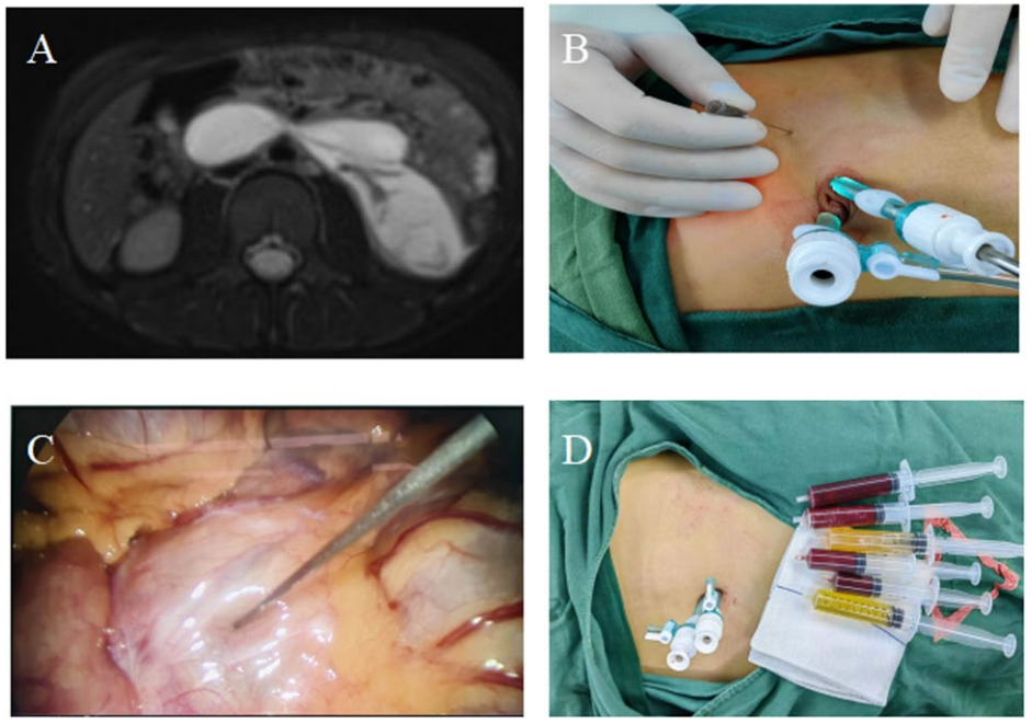
Fig. 1 Key procedural steps of laparoscopic polidocanol foam sclerotherapy (LPFS). A Preoperative planning: MRI (T2-weighted) reveals a septated, high-signal retroperitoneal lymphatic malformation. B Access and targeting: Laparoscopic-assisted puncture is performed, avoiding critical interposed structures. C Visual confirmation: The laparoscopic view provides real-time confirmation of safe needle trajectory and intracystic position. D Diagnostic aspiration: Cyst content is sampled; the presence of both serous and hemorrhagic fluid aids in diagnosis and operative planning
Inclusion criteria: Patients aged <18 years with a confirmed retroperitoneal LMs who underwent LPFS.Exclusion criteria: Patients with lymphatic malformations in other locations requiring a different surgical approach; those with known systemic vascular malformation syndromes; or those with incomplete clinical or follow-up data.
Laparoscopic polidocanol foam sclerotherapy
All procedures were performed under general anesthesia. Pneumoperitoneum was established (maintained at 8–12mmHg) via a 5-mm umbilical trocar. Under laparoscopic guidance, one or two additional working trocars (3-mm or 5-mm) were inserted under laparoscopic guidance. The retroperitoneal lesion was fully exposed and its anatomical relationships were assessed.
A key step involved cyst puncture under direct vision using an 22-gauge Veress needle. Correct intracystic position was rigorously confirmed by aspirating characteristic clear, serous, or chylous lymphatic fluid. If frank blood was aspirated, a critical distinction was made between intracystic hemorrhage and accidental vascular puncture:
1. Intracystic hemorrhage was indicated by the aspiration of dark, non-pulsatile blood that typically ceased after initial drainage, and the cyst wall was observed to collapse.
2. Accidental vascular puncture was suspected if brightred, pulsatile blood was aspirated continuously. In such cases, the needle was immediately withdrawn,pressure was applied laparoscopically, and a newpuncture site was selected.
Onlyafterconfirming an intracystic position was thesclerotherapy proceeded.
Polidocanol foam was prepared intraoperatively using the Tessari method. A three-way stopcock connected two10-mL syringes, one containing 3% polidocanol solution and the other containing room air. With a fixed gas-toliquid ratio of 3:1, the mixture was vigorously agitated through the stopcock for 20 cycles to create a stable, homogeneous foam, which was used within 2 min of preparation to ensure optimal stability.
After maximal aspiration of cystic fluid, the foam was injected slowly under continuous laparoscopic visualization until the cyst wall was fully distended or mild reflux was observed at the puncture site. For multiloculated or septated cysts, individual locules were punctured and treated sequentially to ensure complete sclerosant coverage. The operative field was meticulously inspected for hemostasis and any signs of extracapsular leakage before trocar removal.
Outcome measurements and definitionsClassificationLMs were classified according to the International Society for the Study of Vascular Anomalies (ISSVA) classification system [1, 3].
Efficacy endpoints
Treatment response was assessed by imaging (ultrasound or MRI).
Complete resolution was defined as the reduction of the largest single diameter or dominant cystic component to less than 2 cm, is a well-established criterion for differentiating macrocystic from microcystic lesions, with the former being associated with favorable responses to sclerotherapy [9, 10].
Residual lesion was defined as a persistent cystic structure with a maximum diameter ≥2 cm at the first postoperative follow-up.
Recurrence was defined as the reappearance of a cystic lesion ≥2 cm after documented complete resolution.
Safety endpoints
Complications were monitored throughout follow-up. Specific definitions included.
Ectopic embolism
Clinical symptoms (e.g., acute respiratory distress, neurological deficit) with imaging confirmation.
Adhesive intestinal obstruction
Clinical and radiographic evidence of bowel obstruction requiring intervention.Other complications included allergic reactions, portsite hernia, infection, and intra-abdominal organ injury.
Follow-up protocol
Patients were typically discharged 2–3 days postoperatively once they were tolerating a regular diet and were otherwise asymptomatic. Systematic follow-up was scheduled at 1 month postoperatively with abdominal ultrasound. If a residual lesion measuring ≥2 cm in maximum diameter and/or causing symptoms was identified, a repeat LPFS session was offered. If complete resolution was confirmed at 1 month, a follow-up abdominal MRI was performed at 6 months postoperatively to establish a baseline for long-term assessment. Patients were then advised to undergo annual clinical and ultrasonographic evaluations thereafter to monitor for late recurrence.
Results
Patient characteristics and operative findings
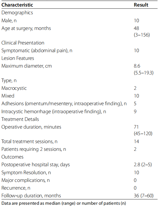
A total of 12 children (10 males and 2 females)with retroperitoneal lymphatic malformations (LMs) were included in this study. Their detailed clinical characteristics are summarized in Table 1. The median age at surgery was 48 months (range: 3–156 months). Ten patients (83.3%) presented with abdominal pain, three of whom also experienced vomiting. The remaining two patients (16.7%)were diagnosed prenatally. All diagnoses were confirmed preoperatively via ultrasonography and magnetic resonance imaging (MRI). Based on MRI findings and the ISSVA classification, there were 2 macrocystic and 10 mixedtype lesions, with a median maximum diameter of 8.6 cm(range: 5.5–19.3 cm). Intraoperatively, intracystic hemorrhage was noted in 9 cases (75%), and adhesions between the cyst and surrounding omentum or mesentery were encountered in 5 cases (41.7%), which required careful laparoscopic adhesiolysis to adequately expose and stabilize the cyst for precise puncture.
Table 1 Clinical Characteristics and Outcomes of 12 Pediatric Patients with Retroperitoneal LMs Treated with LPFS
Treatment efficacy and safety
All procedures were successfully completed under laparoscopy without conversion to open surgery. The key operative steps are illustrated in Fig. 1. The median operative duration was 71 min (range: 45–120 min). All patients achieved complete radiologic resolution. Specifically, 10 patients (83.3%) achieved complete resolution after a single LPFS session. 2 patients (16.7%) with mixed-type lesions had residual cysts (>2 cm) at the first postoperative ultrasound (4 weeks) and subsequently underwent a second LPFS session. Their initial lesion diameters were 8.2 cm and 10.0 cm,with post-procedural residuals measuring 3.2 cm and 3.5 cm, respectively. During the second procedure, dark, old hemorrhagic fluid was aspirated from both cysts. Laparoscopically, the cyst walls appeared slightly thickened and opaque but with clearly discernible boundaries; no dense adhesions or significant tissue plane alterations that could complicate the procedure were encountered. Following the second session, both patients achieved complete radiologic resolution, with final residual lesions measuring less than2 cm. No major complications—such as vascular injury, organ perforation, or clinically significant sclerotherapyrelated adverse events—occurred during the procedures or the immediate postoperative period.
Follow-up outcomes
The median postoperative hospital stay was 2.8 days(range: 2-5days). All patients were followed up postoperatively with a median duration of 36 months (range:7–60 months). Follow-up MRI at 6 months and subsequent annual ultrasound examinations confirmed sustained complete resolution in all cases. Furthermore, all ten patients who presented with preoperative symptoms reported complete and sustained resolution of symptoms following treatment, correlating with the radiologic findings. No recurrence of the lymphatic malformation or long-term complications were observed during the follow-up period.
Discussion
This retrospective study demonstrates that LPFS is a safe and effective minimally invasive approach for the treatment of retroperitoneal LMs in children. In our initial experience with 12 children, the procedure was associated with a high rate of complete resolution with no major complications or recurrence observed over a median follow-up period of 36 months.
Interpretation and comparison
For retroperitoneal LMs located in the deep midline, the therapeutic challenge is particularly pronounced. These lesions are often embedded among critical anatomical structures, including major vessels, the kidneys, and intestinal tracts [11]. Although complete surgical excision has been regarded as the definitive curative treatment with well-established long-term outcomes [4, 6], open surgical excision in this region necessitates extensive dissection within the abdominal and retroperitoneal cavities. This approach not only constitutes considerable invasiveness but also carries a high risk of damaging surrounding tissues [12]. Moreover, even with open exposure, achieving complete resection can be difficult without undue risk. Laparoscopic resection, as reported in series [5], represents a significant minimally invasive advance [13]. However, for lesions intricately adherent to vital retroperitoneal structures, complete dissection can be technically challenging and risks injury [5, 14]. In contrast, LPFS achieved complete radiologic resolution in all cases without the need for open conversion, suggesting it may offer a minimally option for achieving the primary therapeutic goal.Sclerotherapy under imaging guidance is a established minimally invasive option, particularly for unilocular macrocysts. However, for retroperitoneal LMs, especially involving the median of retroperitoneal, the position of is deep, surrounded by many complex anatomical structures, such as intestinal [15, 16], pancreas, spleen[13] and many important vessels. Direct access to these deep-seated lesions through the abdominal wall can be anatomically difficult, and the puncture path may pose a significant risk of injury to interposed abdominal organs. In addition, ultrasound-guided injection of sclerotherapy [15] cannot determine the source of abdominal LMs which is crucial for the selection of treatment methods[14, 17]. The laparoscopic approach employed in our study potentially addresses this limitation. Firstly, laparoscopy provides direct visualization of the lesion and its anatomical relationships, allowing for differential diagnosis of cysts and evaluation of its extent [18]. Secondly, the needle is operated under direct vision not only beneficial for sclerotherapy, but also less likely to damage abdominal organs.The commonly used sclerosing agents for LMs include ing OK-432, bleomycin, doxycycline, and foam of polidocanol or sodium tetradecyl sulfate [1, 19]. A particular limitation of liquid agents is the difficulty in visually distinguishing between injected and uninjected compartments during the procedure, especially in multicystic or septated lesions with spatial overlap. This can lead to incomplete treatment and potential post-procedural residue. To address this issue, we employed polidocanol in a foam formulation. The foam creates a clear and immediate visual contrast within the cyst cavity, allowing for real-time confirmation of distribution (Fig. 2).
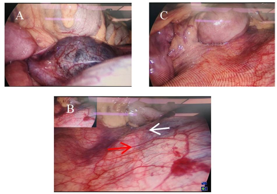
Fig. 2 Figure 2 Intraoperative visualization advantage of foam sclerosant. A Baseline anatomy: Laparoscopic view of the cyst prior to injection, appearing as a single compartment. B Real-time delineation: Upon foam injection, the treated area (red arrow) is immediately and clearly distinguished from the untreated locule (white arrow) by a sharp demarcation line, revealing the true multiloculated anatomy. C Endpoint confirmation: Complete filling of the entire cystic structure with foam, ensuring direct visual confirmation of comprehensive sclerosant distribution and treatment endpoint
This markedly reduces the likelihood of missing a compartment and the need for repeated punctures, thereby potentially enhancing the completeness of ablation and the overall therapeutic efficacy.
Therefore, the favorable preliminary outcomes observed in our series might be attributed to the synergistic combination of laparoscopic visualization and the properties of polidocanol foam [20]. Under laparoscopic direct vision, the surgeon can systematically identify, individually puncture, and ensure complete foam filling of each sizable locule. The foam formulation itself provides an additional advantage, as it fills the cyst, the created visual contrast offers immediate intraoperative feedback, confirming that the entire compartment is treated and leaving little doubt about untreated areas.
Clinical implications and technical recommendations
Notably, intraperitoneal adhesions were encountered in a substantial subset of our patients (5/12, 41.7%). This finding likely reflects the chronic, space-occupying nature of large retroperitoneal LMs, which can incite a local inflammatory response. The presence of adhesions adds a layer of complexity to any surgical approach. In open surgery, dense adhesions increase the risk of visceral injury during dissection. In percutaneous sclerotherapy, they are undetectable and may fix bowel loops in the path of a blind needle, elevating the risk of perforation. The laparoscopic approach directly addresses this challenge. Crucially, the goal of adhesiolysis in LPFS differs fundamentally from that in radical excision [21]. Unlike excision which requires complete mobilization of the entire lesion, LPFS only necessitates a minimal and targeted adhesiolysis to create a safe puncture window and working space. This approach allows for precise needle access while deliberately minimizing unnecessary dissection, thereby reducing the risk of collateral tissue damage and potential bleeding. It is this targeted, rather than exhaustive, dissection that may have contributed to the observed variation in operative duration (range: 45–120 min). This principle underscores a key advantage of LPFS over both open surgery and purely percutaneous techniques: it offers a controlled means to manage anatomical distortion with inherently less tissue trauma. In our series, while all patients ultimately achieved complete resolution, two cases required a second sclerotherapy session, highlighting that managing residual lesions is a key focus for refining LPFS. Our experience suggests that several factors may contribute to residual disease: intracystic hemorrhage, which can dilute the sclerosant and limit endothelial contact [22]; complex multicystic anatomy that hinders uniform foam distribution; and potential foam extravasation through an enlarged or multiple needle tracts. To mitigate these risks, we recommend integrated procedural refinements:for cysts with hemorrhage, gentle irrigation after aspiration may improve sclerosant efficacy; meticulous preoperative MRI review combined with systematic laparoscopic exploration is essential to delineate multicystic structures and reduce missed locales; and operative technique should emphasize minimal needle movement within the cyst, avoiding repeated punctures at the same site to decrease tract size and sclerosant leakage. These technical adjustments, derived from our initial experience, aim to enhance the consistency and completeness of treatment in future applications. However, during the secondary procedures, the residual cysts were easily identifiable laparoscopically with minimal surrounding adhesions, facilitating safe re-puncture and suggesting that the initial foam sclerotherapy does not create a hostile surgical field.Based on the favorable therapeutic outcomes and safety profile preliminarily observed in this study, laparoscopic polidocanol foam sclerotherapy (LPFS) may be considered a viable minimally invasive option for pediatric macrocystic or mixed-type retroperitoneal LMs, particularly those located in anatomically complex regions. To optimize the procedure, our experience highlights several key technical considerations: if the cyst aspiration yields frankly bloody fluid, injection should be paused to exclude accidental vascular puncture; intraoperative observations suggest that minimal foam leakage from the needle tract does not appear to cause clinically significant adhesions, as evidenced in two re-intervention cases, although meticulous technique to minimize leakage remains essential; and the entire procedure requires gentle manipulation, limiting needle movement within the cyst and avoiding repeated punctures at the same site, to prevent tract enlargement, which could lead to sclerosant extravasation and compromised efficacy.
Limitations
Several limitations of this study should be acknowledged. First, the findings are based on a relatively small, singlecenter, retrospective case series, which may limit the statistical power and generalizability of our results. Second, there is an inherent selection bias as all included patients were chosen for surgical intervention. Furthermore, although the mean follow-up period of 36 months is adequate for initial assessment, longer-term follow-up would provide more definitive evidence regarding sustained efficacy and absence of late recurrence. Finally, the lack of a direct control group precludes comparative effectiveness conclusions.
Conclusion
In conclusion, this study suggests that laparoscopic polidocanol foam sclerotherapy (LPFS) is a safe and effective minimally invasive treatment for pediatric retroperitoneal LMs within our cohort. To validate and extend these promising preliminary results, future research should prioritize conducting larger-scale, multicenter, prospective studies, ideally with randomized controlled designs that directly compare LPFS to standard treatment options. Such studies should aim to enroll more patients, incorporate longer follow-up durations, and further refine the optimal indications for this technique, such as specific cyst sizes, morphological classifications, and anatomical locations.
Abbreviations
LMs Lymphatic malformations
LPFS Laparoscopic polidocanol foam sclerotherapy
ISSVA The International Society for the Study of Vascular Anomaly
Acknowledgements
None.
Authors’contributions
JB, SL and YL conceived and designed the study, acquired the data, drafted the manuscript, and performed the analysis and interpretation. ZB andYF critically revised the manuscript. All authors reviewed the final version of the manuscript.
Funding
Startup Fund for scientific research, Fujian Medical University (Grant number:2022QH1224 and 2024QH1253). Fujian Provincial Natural Science Foundation(Grant number: 2024J011133). Medicine Research Project of Fujian Children’sHospital (Grant number: ETK2025007).
Data availability
The raw data supporting the conclusions of this article will be made available by the authors, without undue reservation.
Declarations
Ethics approval and consent to participate
The study was approved by the Ethics Committee of Fujian Children’sHospital(2023ETKLR05065) based on the principles of the Declaration of Helsinki and written informed consent has been obtained from the children and their parents.
Consent for publication
Not applicable.
Competing interests
The authors declare no competing interests.