发布时间:2026-10-30 08:49:08 来源: 浏览次数:55
《DERMATOLOGIC SURGERY》2026;00:1–2
Cannula-Assisted Sclerotherapy for Axillary Osmidrosis
Wenhui Liu, MD, PhD*† Dingrong Gong, MD† Yu Wang, MD†Jingxia Zhang, MD**Department of Plastic Surgery, The First Affiliated Hospital of Zhengzhou University, Zhengzhou, Henan, China;†Department of Aesthetic Medicine, Hotan District People’sHospital, Xinjiang, China
http://dx.doi.org/10.1097/DSS.0000000000005431
Sclerotherapy for axillary osmidrosis (AO) can be performed with a cannula instead of a needle, a technique that potentially enhances safety,improves efficacy, and increases patient comfort. To the best of our knowledge, this is the first report of cannulaassisted sclerotherapy for AO. AO is a distressing condition characterized by malodor that imposes significant social pressure on patients. Numerous treatments have been developed for AO, with sclerotherapy emerging as a promising option. 1,2Some physicians recommend using needle for the injection of sclerosing agents such as polidocanol. However, in our practice, we have found that using a cannula instead of a needle serves this purpose better.
Technique
The technique we propose for sclerotherapy of AO with a cannula is quite similar to the injection of dermal fillers with a cannula. Compound lidocaine ointment should be applied before injection. After the required waiting time has elapsed, the ointment is removed, and the area to be injected is marked and sterilized (Figure 1A) . A port is then made with a needle thicker than the cannula (Figure 1B) . The cannula is subsequently introduced through this port, and blunt dissection should be thoroughly performed within the target injection area (Figure 1C). Finally, injection should be made slowly and gently, stopping right at the border of the target area (Figure 1D) . For experienced physicians, injection can also be performed during the blunt dissection process. A practical point is that the cannula tip should always be palpated with the fingertips. The tip should be oriented upward rather than downward during injection to prevent penetration into deeper layers. All other parameters, such as drug concentration and dressing protocol, remain identical to those used in needle-based procedures. Two videos are presented to illustrate this technique (see Supplemental Digital Content, Video 1, https://links.lww.com/DSS/C53 and Video 2, https://links.lww.com/ DSS/C54 ) .
Discussion
Histological studies show that axillary apocrine glands are confined mainly to the deep reticular dermis and superficial subcutaneous layer, whereas major vessels and nerves course deeper beneath the fascia or at the muscle surface.3The blunt tip of a cannula therefore dissects naturally along this relatively loose and avascular subdermal plane, keeping the sclerosing agent within the gland-bearing layer and away from major vessels. This anatomical alignment may reduce the risk of intravascular injection, skin necrosis, and neurovascular injury compared with a sharp needle.
The safety profile may be enhanced by the blunt tip, which mitigates inadvertent intradermal deposition and vascular injury. The greater length of the cannula shaft permits the entry port to be situated farther from the treatment boundary, potentially reducing leakage-related complications.
Efficacy may be optimized through blunt dissection, which creates a broader and more uniform subdermal plane,4,5 facilitating homogeneous drug dispersion and a consistent fibrotic response. In contrast, a sharp needle deposits the agent in a more focal and irregular manner, which may result in uneven distribution and inconsistent fibrotic response.
Patient comfort and procedural efficiency may also improve because the extended reach of the cannula allows a larger treatment area to be covered through fewer puncture sites, reducing pain, shortening procedure duration, and streamlining injection.
The cannula may be particularly suitable for novice practitioners, who gain a greater margin of safety, and for physicians experienced in dermal filler injection, who can adopt the technique with minimal additional training. Even without prior filler experience, the technique remains intuitive and straightforward.
Nevertheless, cannulas are not without limitations. They are generally softer than needles and may lack sufficient rigidity for effective blunt dissection in patients with axillary scarring from previous treatments. They are also more costly and not universally available. Under such circumstances, needles remain necessary, and longer needles are preferable to shorter ones. Cannulas are not entirely riskfree; blood vessel wall penetration and ischemic events remain possible with inappropriate plane selection or excessive injection pressure. The potential advantages described here are based on single-center clinical experience and have not been quantified in controlled studies. Largesample randomized trials are warranted to compare cannula and needle techniques.
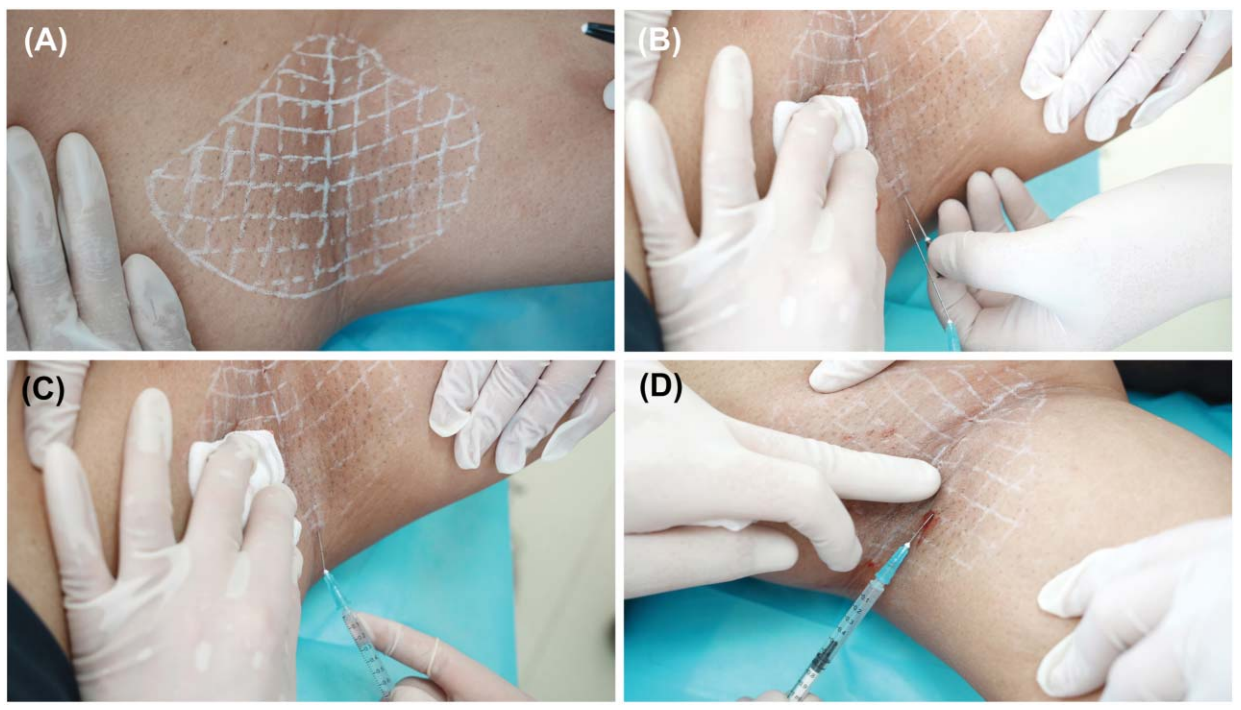
Figure 1. Cannula-assisted sclerotherapy for axillary osmidrosis. (A) Marking and sterilization. (B) Creation of a port for cannula introduction. (C) Blunt dissection with the cannula. (D) Injection of the sclerosing agent.
Conclusion
When sclerotherapy is indicated for AO, the cannula represents a reasonable and potentially preferable alternative to the needle in appropriately selected patients. Future well-designed randomized trials are needed to compare these 2 techniques.
作者:刘文辉,医学博士,郑州大学第一附属医院援疆专家;现任和田地区人民医院烧伤整形科主任、医学科主任


